Meet our Doctor
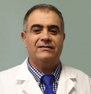
From your very first office visit with Dr. Elie C. Daniel, you’ll know your foot and ankle health is under the most capable and compassionate care. He is friendly, genuinely concerned, and ready to listen to you and answers any questions you may have. He will take the time to thoroughly examine you and use state-of-the-art diagnostics to help determine and correct the underlying problem—and not just treat your symptoms.
When making his diagnosis, Dr. Daniel considers the whole body and how biomechanics—the mechanics of human movement—factors into your feet or ankle condition. He’ll explain all of this to you so you can be knowledgeable, confident, and involved in your own treatment and recovery.
Dr. Daniel has over 30 years of experience treating general podiatric and diabetic foot conditions. He always looks for conservative methods to alleviate foot and ankle problems. However, if surgical intervention is necessary to achieve the best outcome for you, Dr. Daniel and his team will keep you informed throughout the process—from evaluation and surgical procedure to post-operative care.
Dr. Daniel specializes in HyProCure, diabetic limb salvage, correcting bony misalignments, restoring range of motion, and treating ankle injuries. He is also the leader in ankle reconstructive surgery, performing implants and transplants for his patients—including the very first ankle replacement and ankle transplant in his area.
If you are having any type of foot or ankle pain, discomfort, or decrease in mobility, Dr. Daniel can help you. Both he and his entire team look forward to getting you back to your normal activities as soon as possible so you can live life to the fullest.
Meet our Doctor
A well-defined, corticated, bipartite ossicle embedded within the substance of the plantar fascia at the plantar midfoot is described. The structure was identified as an incidental finding on weight-bearing plain radiographs in an asymptomatic 56-year-old female. Serial imaging spanning five years documented progressive maturation from two faint mineralization foci to a fully formed corticated ossicle, supporting a primary developmental etiology. Digital grayscale pixel intensity analysis confirmed greater radiographic density of the ossicle (mean 45,748 US units) relative to the surrounding plantar fascia (mean 43,953 US units), consistent with organized osseous tissue. Given its mid-plantar location with no adjacent bone of origin, the structure most closely resembles a sesamoid-type ossicle developing within a fibrous structure. The provisional designation Os Danielis (os plantare fasciae midpedis) is proposed. Its intrafascial position may create a focal zone of reduced tensile capacity — a locus minoris resistentiae — analogous to the mechanical vulnerability of the os tibiale externum at the posterior tibial tendon insertion. Clinicians should be aware of Os Danielis in the differential diagnosis of medial plantar midfoot pain.
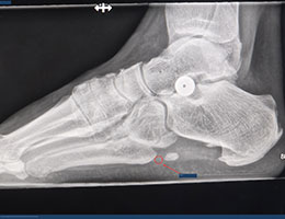
Figure 1.
Lateral oblique weight-bearing radiograph of the right foot (2026) demonstrating Os Danielis on the plantar midfoot (circle and arrow). The ossicle demonstrates a well-corticated rim surrounding a central radiolucency consistent with a medullary cavity — the characteristic bullseye appearance. The sinus tarsi implant is noted in satisfactory position in the sinus tarsi; its presence is incidental and bears no relationship to Os Danielis.
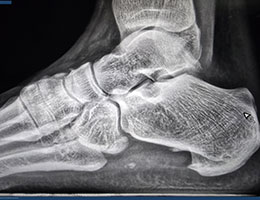
Figure 2.
Lateral radiograph of the right foot (2021, early stage) showing two faint, poorly corticated mineralization foci in the plantar midfoot soft tissue, representing the earliest documented stage of Os Danielis development. No sinus tarsi implant is present in this image, confirming that the ossicle predates any subsequent intervention and is entirely independent.
Sesamoid bones develop embedded within tendons or fibrous soft tissue structures, unconnected to any adjacent bone, and serve to modify the direction of mechanical forces within the structure in which they are embedded (Keles-Celik et al. 2017). Accessory ossicles, by contrast, represent unfused secondary ossification centers of adjacent bones. The plantar fascia is a dense fibrous aponeurosis originating at the medial calcaneal tubercle and extending distally as five fiber bundles toward the forefoot (Vanhoenacker et al. 2014). Its mechanical integrity relies upon the continuity of its parallel-oriented type I collagen fiber network (Bolgla and Malone 2004).
We report an incidental finding of a previously undescribed ossicle within the substance of the plantar fascia at the plantar midfoot, supported by five-year serial radiographic documentation and digital grayscale density analysis. The provisional eponymous designation Os Danielis (os plantare fasciae midpedis) is proposed for this structure.
A 56-year-old female presented for routine postoperative radiographic evaluation following extra-osseous talotarsal stabilization (EOTTS) sinus tarsi implantation for recurrent dislocation of the talotarsal joints (Graham et al. 2011). She reported no pain, tenderness, or functional limitation in the plantar midfoot region. Medical history was unremarkable for inflammatory arthropathy, metabolic bone disease, connective tissue disorder, or prior plantar foot trauma.
Physical examination revealed no palpable nodule, focal tenderness, induration, or soft tissue abnormality at the ossicle site. Systematic bimanual palpation with passive digital extension to engage the windlass mechanism elicited no discomfort. Gait analysis and neurovascular examination were within normal limits.
Weight-bearing plain radiographs of the right foot in lateral, lateral oblique, and axial projections revealed an incidental well-defined osseous structure on the plantar aspect of the midfoot, projecting within the expected course of the medial band of the central plantar fascia, inferior to the naviculocuneiform articulation (Fig. 1). The ossicle demonstrated:
Digital grayscale pixel intensity measurements using the PACS region-of-interest (ROI) tool yielded ossicle values of 45,075 and 46,420 US units (mean 45,748 US) versus surrounding plantar fascial tissue values of 43,867 and 43,996 US units (mean 43,953 US) — a difference of approximately 4.1%, consistent with greater mineral density of organized osseous tissue relative to dense fibrous connective tissue.
Archival radiographs from five years prior confirmed the pre-existing and independent nature of this ossicle (Fig. 2). Two discrete, faint, poorly corticated mineralization foci were identified in 2021, with progressive coalescence and cortical formation documented through to the fully formed mature bipartite ossicle of 2026. The absence of any precipitating trauma, surgery, or inflammatory event during this period argues against an acquired or heterotopic etiology.
Embryological Basis
Sesamoid bones arise through endochondral ossification within fibrous or tendinous structures subjected to repetitive mechanical compression and tension during development. The plantar fascia, as a principal tension-bearing structure of the foot, is subject to substantial repetitive compressive and tensile forces from the earliest stages of weight-bearing ambulation. It is postulated that Os Danielis represents a focal center of endochondral ossification that arose within the medial band of the central plantar fascia in response to the localized mechanical stress environment at the plantar midfoot. This developmental mechanism is analogous to that proposed for sesamoid bones within the flexor hallucis brevis and other fibrous plantar structures, in which repetitive focal loading during gait triggers chondrification and subsequent ossification of mesenchymal tissue within the fibrous matrix (Keles-Celik et al. 2017). The five-year longitudinal imaging series documenting the progressive maturation of Os Danielis from discrete mineralization foci to a fully corticated ossicle is consistent with this primary endochondral developmental process.
Classification
The absence of any adjacent bone of origin on all radiographic projections argues against the accessory ossicle classification. Os Danielis more closely satisfies the definition of a sesamoid bone — a bone embedded within a fibrous or tendinous structure, unconnected to any adjacent bone (Keles-Celik et al. 2017). We propose it be classified as a sesamoid-type ossicle of the plantar fascia, pending histological confirmation.
Differential Diagnosis
Several entities must be considered in the differential diagnosis of a plantar midfoot osseous density (Table 1). Plantar fascia enthesopathy presents proximally at the calcaneal insertion with a spiculated morphology and no medullary cavity. Plantar fibromatosis presents as an isoechoic fascial nodule without bony architecture on ultrasound (Tomac et al. 2023). The Lisfranc ligament complex is a deep periarticular structure at the tarsometatarsal joint level, significantly deeper than Os Danielis, and Lisfranc pathology is fundamentally articular in nature (Weatherford et al. 2025). Dystrophic calcification produces amorphous mineral deposits without organized cortical architecture. Heterotopic ossification follows a characteristic zonal maturation pattern associated with trauma or surgery — not present in this case.
Table 1. Differential diagnosis of plantar midfoot osseous density and distinguishing features from Os Danielis.
| Differential Diagnosis | Key Feature | Distinguishing from Os Danielis |
|---|---|---|
| Plantar enthesopathy | Spiculated, no cortex | Proximal calcaneal; no medullary cavity |
| Plantar fibromatosis | Isoechoic on US; no calcification | No bony architecture |
| Lisfranc ligament pain | Deep periarticular; diastasis on X-ray | Deeper; joint-based pathology |
| Dystrophic calcification | Amorphous; no cortex | No organized cortex or medullary cavity |
| Heterotopic ossification | Zonal peripheral pattern | Trauma history; peripheral-to-central |
Biomechanical Implications and Clinical Significance
The intrafascial position of Os Danielis displaces and interrupts the organized collagen fiber network of the plantar fascia, potentially creating a focal zone of reduced tensile capacity — a locus minoris resistentiae. This concept finds a useful clinical parallel in the os tibiale externum, where the ossicle creates a mechanically deficient posterior tibial tendon insertion predisposing to tendon dysfunction and failure (Romanowski and Barrington 1992). Similarly, Os Danielis may represent a site of focal biomechanical vulnerability within the plantar fascia.
In view of the asymptomatic presentation, advanced imaging with CT and MRI is not being pursued currently and is reserved as a contingency should symptoms develop. CT would characterize the cortical architecture and three-dimensional morphology; MRI would assess fatty marrow signal on T1-weighted sequences — the definitive criterion distinguishing a true ossicle from calcification. Custom functional foot orthoses providing triplanar arch support with a plantar fascial groove accommodation beneath the ossicle site are recommended (Landorf et al. 2006). Corticosteroid injection near the site warrants caution given the documented risk of fascial rupture (Acevedo and Beskin 1998) and the additional weakening effect of steroid on collagen (Kennedy and Willis 1976). Serial weight-bearing radiographs at twelve-month intervals are recommended to monitor for interval change.
Os Danielis (os plantare fasciae midpedis) represents a potentially novel sesamoid-like ossicle embedded within the medial band of the central plantar fascia at the plantar midfoot. Its organized cortical architecture, bipartite morphology, radiographic density greater than the surrounding fascia, and five-year developmental maturation sequence serve to distinguish it from pathological calcification or heterotopic ossification. Its mid-plantar location with no adjacent bone of origin most closely satisfies the definition of a sesamoid-type ossicle within a fibrous structure. Clinicians should be aware of this entity in the differential diagnosis of medial plantar midfoot pain. Future studies examining prevalence and histological characterization will further clarify its classification and clinical significance.
Conflict of interest: The author declares no conflicts of interest.
Funding: No funding was received for this work from any public, commercial, or not-for-profit organization.
Ethics and patient consent: Written informed consent was obtained from the patient for publication of this case report and all associated images in accordance with HIPAA regulations. The signed consent form is retained in the patient's medical record and is available for review upon request.
Artificial intelligence disclosure: Claude (Anthropic, PBC; claude.ai), an artificial intelligence language model, was used as a writing assistance tool during manuscript preparation. All clinical observations, radiographic findings, data, and intellectual content are entirely those of the author, who takes full responsibility for the accuracy and integrity of all content.
No competing interests reported.




Dr. Daniel and the staff are amazing! I appreciate the kindness and the results.
Dr. Elie Daniel did a bunion correction lapiplasty surgery on me and he and his staff were so caring, no matter what questions or concerns I had he got right back to me himself. I would recommend Dr. Elie Daniel, he will be doing my other foot and I feel I’m in great hands!
I highly recommend Dr. Daniel. He genuinely cares about his patients. His professionalism and knowledge make him very trustworthy.
I had a bone spur removed from my left big toe and it looks very good and it has healed up very nicely. Dr. Daniel and his office staff are very kind and considerate and will answer any questions or concerns you may have. Their office is always very neat and clean, and I totally recommend this podiatrist's office they have been very good to me!!!
Dr. Daniel uses the latest technology for your injuries to heal faster and better. He also has a YouTube channel: Dr. Elie Daniel for more educational content.
This practice uses the latest technology and cares about their patients.
Dr. Daniels has made amazing accomplishments in regard to my ankle, unlike others. One of the best doctors I have met. In my book, he is royalty in the medical field. I would highly recommend Dr. Daniels for your foot and ankle needs.
Dr. Daniel is such a caring and amazing doctor. Took time to help me with a prescription while he was at home. Took time to correspond with both myself and the pharmacy just to help me due to the pain I was in. Wish all Doctors cared for their patients as he does.
Dr. Daniel is always a pleasure and you can tell he genuinely cares about helping his patients. Most doctors will just do the minimum and send you on your way when they can’t see an obvious problem, but Dr.Daniel searches for the answer and does everything in his power to help you get better. 10/10 would recommend.
Connect With Us

 Elie C. Daniel, DPM